No products in the cart.
U.S. FILLED & FINISHED | ≥99% COA-VERIFIED PURITY
LC-MS VERIFIED IDENTITY | INDEPENDENTLY TESTED
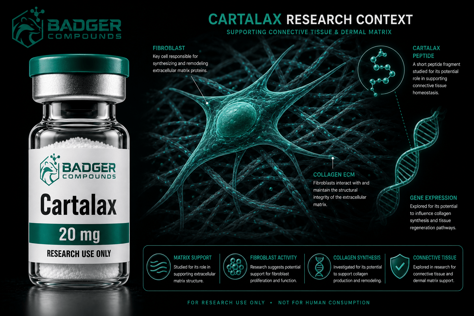
Cartalax peptide research centers on the AED tripeptide, a Khavinson short-peptide bioregulator studied across connective tissue and cellular aging models, including fibroblast function, extracellular matrix remodeling, senescence markers, and its role as a component of a cartilage-derived complex investigated in osteoarthritis.
Cartalax Peptide Research: The AED Short Peptide Bioregulator
Cartalax is a synthetic tripeptide (Ala-Glu-Asp, AED) developed within the Khavinson short-peptide bioregulator program. Preclinical investigations have examined it across connective tissue, cellular aging, and cartilage research contexts, spanning fibroblast function, matrix remodeling, senescence markers, and gene expression.
What Is Cartalax?
Cartalax is an ultrashort synthetic tripeptide composed of three amino acids, alanine, glutamic acid, and aspartic acid, in the sequence Ala-Glu-Asp (AED). It has a molecular weight of approximately 333.29 Da and the molecular formula C12H19N3O8. It belongs to the family of short regulatory peptides developed at the St. Petersburg Institute of Bioregulation and Gerontology under the research program associated with Professor Vladimir Khavinson.
A point of accuracy worth noting: some suppliers list Cartalax as a four-amino-acid tetrapeptide, but the compound indexed in the scientific literature is the three-residue AED tripeptide. Notably, AED shares its full sequence with the first three residues of Epitalon (Ala-Glu-Asp-Gly), and the two peptides are studied together in several of the same papers. For the broader geroprotective context of that peptide family, see our overview of Epitalon and cellular aging research.
The Khavinson Short Peptide Concept
The research framework behind Cartalax is the bioregulator hypothesis developed over decades by the Khavinson group. The central proposal is that very short peptides, typically two to four amino acids, can act as tissue-specific regulators of gene expression rather than as receptor-binding signaling molecules in the conventional sense. In this model, the peptides are small enough to enter cells and the nucleus, where they are proposed to interact with DNA and influence the transcription of specific genes.
This is a mechanistic hypothesis under active investigation, not a settled pathway. In one computational and cell-based study, Khavinson and colleagues (2014) constructed models suggesting that AED and a related peptide form energetically favorable complexes with particular DNA sequences in the minor groove, and correlated this with changes in the expression of aging-associated genes in renal cell cultures. The DNA-interaction mechanism should be understood as a proposed model supported by preliminary data rather than a fully established mechanism of action.
Research framing: All findings referenced in this post derive from preclinical cell-based and animal research models, published largely by a single research group. Cartalax is supplied by Badger Compounds for laboratory research use only and is not intended for human or veterinary use. No therapeutic conclusions should be drawn from the research discussed here.
Fibroblast and Connective Tissue Research
The most directly relevant connective tissue work is a study by Lin’kova and colleagues (2016) in the Bulletin of Experimental Biology and Medicine, which examined short peptides including AED in cultured skin fibroblasts undergoing replicative aging. Using immunofluorescent confocal microscopy, the researchers assessed markers of proliferation (Ki-67), regeneration and aging (CD98hc), apoptosis (caspase-3), and extracellular matrix remodeling (MMP-9).
The reported findings are relevant to connective tissue biology because fibroblasts are the principal matrix-producing cells of connective tissue. All the studied peptides inhibited synthesis of MMP-9, a matrix metalloproteinase whose activity increases during fibroblast aging and contributes to matrix breakdown, and enhanced expression of Ki-67 and CD98hc, which decline as cells age. AED specifically, along with the AEDG peptide, was reported to suppress caspase-dependent apoptosis that rises during the aging of these cell cultures. These are the observations that most directly underpin the connective tissue research framing for AED.
Matrix Remodeling
In aging skin fibroblasts, short peptides including AED were reported to inhibit MMP-9, a matrix metalloproteinase associated with age-related connective tissue breakdown.
Proliferation Markers
The same research associated peptide treatment with enhanced Ki-67 and CD98hc expression, markers that ordinarily decline during fibroblast aging in culture.
Apoptosis Modulation
AED and the related AEDG peptide were reported to suppress caspase-dependent apoptosis that increases during the aging of fibroblast cultures.
Cellular Aging and Gene Expression
Beyond fibroblasts, AED has been examined in other cellular aging systems. Ashapkin and colleagues (2020), publishing in Molecular Biology Reports, studied AED alongside the peptides KED and KE in human mesenchymal stem cells aged in culture. The research reported that these peptides at nanomolar concentrations modulated the expression of several genes involved in cell aging, including IGF1, FOXO1, TERT, and NFkB, with the peptides enhancing IGF1 expression several-fold across the aging models tested.
In parallel renal work, Chalisova and colleagues (2015) reported that AED, designated T-31 in that study, stimulated proliferation and reduced apoptosis in kidney tissue cultures from both young and old animals, though to a lesser degree than a whole polypeptide complex. The 2014 renal-culture study noted earlier connected AED treatment to decreased expression of the senescence markers p16, p21, and p53 and increased expression of SIRT-6. Taken together, these studies position AED as a short peptide associated with modulation of senescence-related gene expression across several cell types, with connective tissue fibroblasts being the most directly relevant to its research profile.
| Research Model | System | Reported Observation |
|---|---|---|
| Skin fibroblast aging | Cultured fibroblasts (Lin’kova 2016) | Inhibited MMP-9; raised Ki-67 and CD98hc; AED suppressed apoptosis |
| Mesenchymal stem cell aging | FetMSC cultures (Ashapkin 2020) | Modulated IGF1, FOXO1, TERT, NFkB expression at nanomolar levels |
| Renal cell aging | Renal cell culture (Khavinson 2014) | Decreased p16, p21, p53; increased SIRT-6; proposed DNA interaction |
| Kidney tissue renewal | Young and old rat explants (Chalisova 2015) | Stimulated proliferation, reduced apoptosis versus control |
| Neuronal differentiation | Periodontal ligament stem cells (Caputi 2019) | AED studied within a peptide compound influencing differentiation markers |
Cartilage and Osteoarthritis Research Context
AED also appears in the cartilage research literature, though in a specific and qualified way worth stating precisely. A 2023 review by Lin’kova and colleagues in the International Journal of Molecular Sciences, focused on peptide regulation of chondrogenic stem cell differentiation, notes that AED is one of the short peptides contained in a polypeptide complex (PCC) isolated from animal cartilage tissue. That complex, developed at the St. Petersburg Institute of Bioregulation and Gerontology, is described as a regulator of cartilage and bone tissue repair and is being investigated as a chondroprotector in models of osteoarthritis.
Importantly, the review does not present AED itself as a demonstrated driver of cartilage matrix synthesis. Rather, it restates AED’s documented gene-expression effects from mesenchymal stem cell aging studies, its influence on NFkB, IGF1, and TNKS2, and offers these as a basis for hypotheses about possible relevance to osteoarthritis-associated inflammation and tissue maintenance. The peptides the same review identifies as most promising for directly driving chondrogenic differentiation are a separate set of growth-factor-based and matrix-mimetic sequences. AED’s connection to cartilage research is therefore best described as its inclusion in a cartilage-derived peptide complex together with a set of mechanistic hypotheses, not as established chondrogenic activity.
Cartalax is a research-use-only peptide. Badger Compounds supplies Cartalax exclusively for qualified research applications in controlled laboratory settings. The research summarized here does not establish safety or efficacy in any human population.
Scope and Limitations of the Evidence
Responsible use of Cartalax as a research tool requires an accurate picture of how thin and specific the evidence base actually is, and this is where much of the available commentary overstates the case. The published literature on AED is small, consists of a handful of studies, and originates almost entirely from a single research program. Several of the studies are published in specialized gerontology journals, and some primary reports are in Russian. In most experiments, AED is not studied in isolation but as one of several short peptides tested together, which can make it difficult to attribute a specific effect to AED alone.
The tissue-specificity central to the bioregulator hypothesis also cuts both ways. Not every model shows activity: in a study of thymocyte aging, AED, again designated T-31, did not produce the geroprotective effect that a different peptide in the same experiment did. This is a useful reminder that results in one cell type do not automatically transfer to another. On cartilage specifically, no primary study has demonstrated AED alone driving cartilage matrix synthesis or chondrocyte differentiation; its documented link to that field is its inclusion in a cartilage-derived polypeptide complex studied for osteoarthritis, together with mechanistic hypotheses drawn from its gene-expression effects. No human clinical data exists for this compound. Cartalax is best understood as an early-stage research peptide for probing short-peptide effects on cellular aging and connective tissue biology, not as a validated intervention for any outcome.
Single-Program Literature
Most published AED research originates from one research group, and the peptide is frequently studied as part of a mixture rather than in isolation.
Preclinical and Model-Dependent
All evidence is from cell and tissue models, effects vary by cell type, and no human clinical data has been reported for this compound.
Research Pathways Associated with Cartalax
Preclinical literature has examined AED and related short peptides across several interconnected research contexts:
Fibroblast proliferation (Ki-67) Extracellular matrix remodeling (MMP-9) Caspase-dependent apoptosis Cellular senescence markers (p16, p21, p53) SIRT-6 and IGF1 expression Proposed peptide-DNA interaction Chondroprotective polypeptide complex (OA) Short-peptide bioregulationCartalax – Research Grade, ≥99% (HPLC)
Badger Compounds supplies Cartalax as a lyophilized research peptide, six-round independently tested per batch with publicly viewable COAs. For qualified laboratory research use only.
View Cartalax- Lin’kova NS, Drobintseva AO, Orlova OA, et al. Peptide regulation of skin fibroblast functions during their aging in vitro. Bull Exp Biol Med. 2016;161(1):175-178. PMID 27259496
- Ashapkin V, Khavinson V, Shilovsky G, Linkova N, Vanuyshin B. Gene expression in human mesenchymal stem cell aging cultures: modulation by short peptides. Mol Biol Rep. 2020;47(6):4323-4329. PMID 32399807
- Khavinson VKh, Tarnovskaia SI, Lin’kova NS, et al. Tripeptides slow down aging process in renal cell culture. Adv Gerontol. 2014;27(4):651-656. PMID 25946838
- Chalisova NI, Lin’kova NS, Nichik TE, et al. Peptide regulation of cells renewal processes in kidney tissue cultures from young and old animals. Bull Exp Biol Med. 2015;159(1):124-127. PMID 26033601
- Caputi S, Trubiani O, Sinjari B, et al. Effect of short peptides on neuronal differentiation of stem cells. Int J Immunopathol Pharmacol. 2019;33:2058738419828613. PMID 30791821
- Lin’kova NS, Polyakova VO, Trofimov AV, Kvetnoy IM, Khavinson VKh. Peptidergic regulation of thymocyte differentiation, proliferation, and apoptosis during aging of the thymus. Bull Exp Biol Med. 2011;151(2):239-242. PMID 22238759
- Linkova N, Khavinson V, Diatlova A, Myakisheva S, Ryzhak G. Peptide regulation of chondrogenic stem cell differentiation. Int J Mol Sci. 2023;24(9):8415. PMID 37176122
Disclaimer: Cartalax is supplied by Badger Compounds for laboratory and in vitro research use only. It is not intended for human or veterinary use, and no product sold by Badger Compounds is approved for therapeutic, diagnostic, or clinical application. All research findings referenced in this post are derived from preclinical cell-based and animal model studies. No clinical efficacy or safety conclusions should be drawn from the data summarized here.