No products in the cart.
U.S. FILLED & FINISHED | ≥99% COA-VERIFIED PURITY
LC-MS VERIFIED IDENTITY | INDEPENDENTLY TESTED
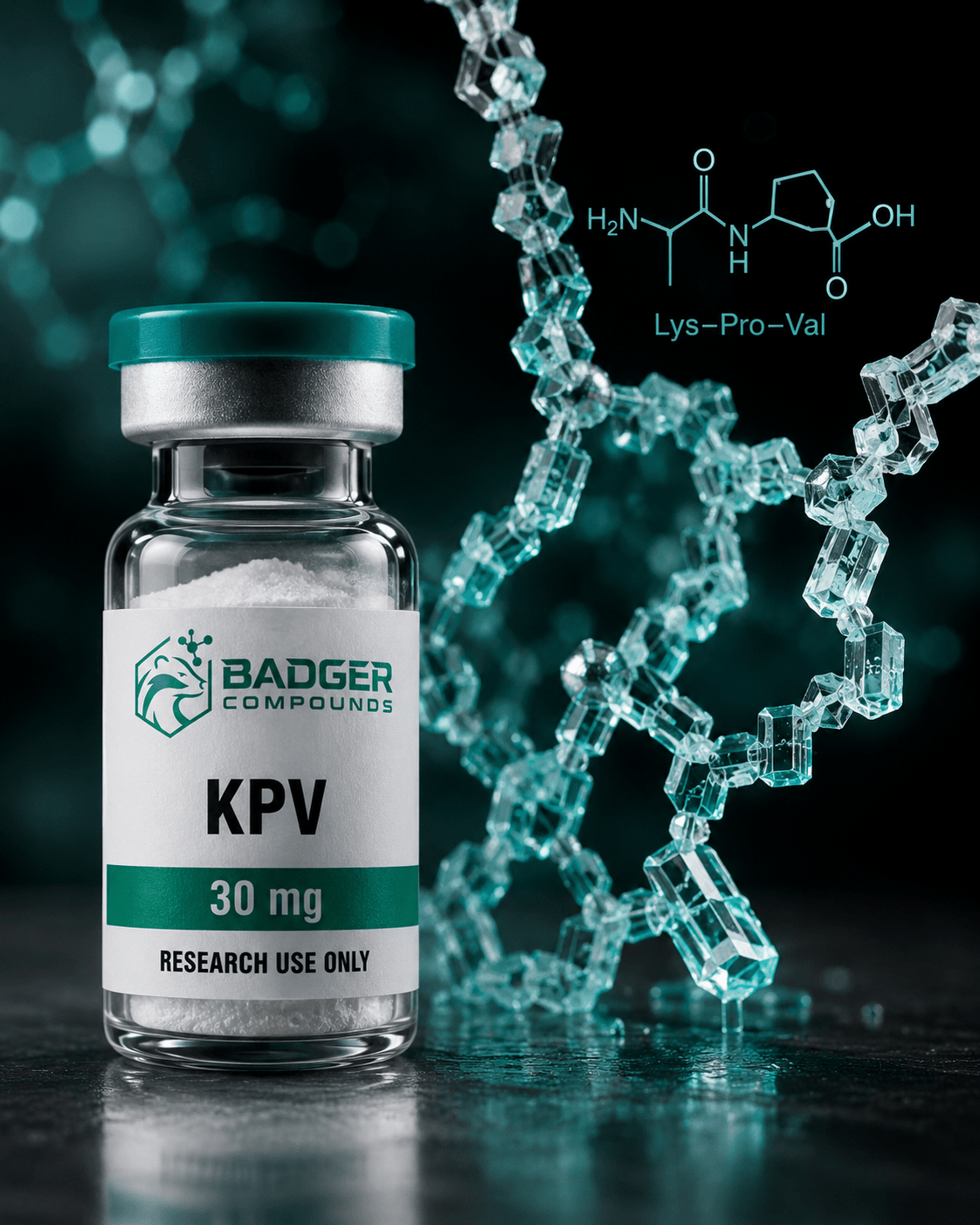
KPV peptide anti-inflammatory research centers on a naturally occurring tripeptide derived from the C-terminal sequence of alpha-melanocyte-stimulating hormone, where preclinical studies have investigated its capacity to modulate NF-kB signaling independent of classical melanocortin receptor engagement.
KPV Peptide Anti-Inflammatory Research: The Alpha-MSH Tripeptide
KPV (Lys-Pro-Val) is the C-terminal tripeptide of alpha-melanocyte-stimulating hormone. Preclinical investigations have characterized its capacity to attenuate NF-kB and MAP-kinase inflammatory cascades through a largely receptor-independent mechanism, making it a structurally minimal subject of ongoing research interest.
What Is KPV?
Alpha-melanocyte-stimulating hormone (alpha-MSH) is a 13-amino-acid neuropeptide processed from the proopiomelanocortin (POMC) precursor. Its C-terminal sequence, positions 11 through 13, is the tripeptide Lys-Pro-Val, designated KPV. Researchers identified KPV as a minimal structural unit that retains meaningful anti-inflammatory activity in preclinical models while lacking the pigmentary side-effect profile associated with the full-length hormone, a property that has sustained interest in the tripeptide across multiple research disciplines.
KPV is also studied as a component of multi-peptide research blends alongside compounds such as GHK-Cu, the copper tripeptide investigated in skin biology research, where co-formulation approaches are of interest in tissue and inflammatory research contexts.
Structural Origins in the Melanocortin System
The melanocortin peptide family, which includes alpha-MSH, beta-MSH, and gamma-MSH, signals primarily through five G-protein-coupled melanocortin receptor subtypes (MC1R through MC5R). The canonical anti-inflammatory pharmacophore of alpha-MSH is generally considered to reside in its core sequence (His-Phe-Arg-Trp at positions 6-9), which binds melanocortin receptors and elevates cyclic AMP. KPV, by contrast, lacks this core binding motif entirely.
Preclinical dissection studies reported by Getting and colleagues demonstrated that KPV’s anti-inflammatory activity in crystal-induced peritonitis models was not blocked by the MC3/4-receptor antagonist SHU9119 and was preserved in recessive-yellow mice lacking functional MC1R, observations consistent with a mechanism that diverges from classical melanocortin receptor engagement. Brzoska and colleagues subsequently reviewed this body of evidence and noted that KPV retains broad anti-inflammatory capacity in research models despite the absence of the receptor-binding pharmacophore.
Research framing: All findings referenced in this post derive from preclinical cell-based and animal research models. KPV is supplied by Badger Compounds for laboratory research use only and is not intended for human or veterinary use. No therapeutic or clinical conclusions should be drawn from the preclinical data discussed here.
NF-kB Inhibition and PepT1-Mediated Cellular Uptake
A foundational mechanistic study by Dalmasso and colleagues, published in Gastroenterology (2008), investigated how KPV exerts anti-inflammatory effects in intestinal epithelial and immune cell models. The research found that nanomolar concentrations of KPV inhibit activation of the NF-kB and MAP-kinase inflammatory signaling pathways and reduce secretion of pro-inflammatory cytokines including IL-1beta, IL-6, TNF-alpha, and IL-8.
Crucially, the study identified PepT1, the di/tripeptide transporter ordinarily expressed in the small intestine and upregulated in colonic tissue under inflammatory conditions, as the cellular entry mechanism for KPV in both intestinal epithelial cells and immune cells. Blocking PepT1 abolished KPV’s anti-inflammatory effects, establishing transporter-mediated uptake as a key step in the observed pathway. In murine colitis models (DSS-induced and TNBS-induced), KPV administration was associated with reduced histological inflammation markers and decreased pro-inflammatory cytokine expression at the mRNA level.
NF-kB Suppression
Preclinical investigations report KPV inhibits IkB-alpha degradation, stabilizing the inhibitory complex and reducing nuclear translocation of the p65 RelA subunit in epithelial and immune cell models.
MAP-Kinase Pathway
Research models indicate concurrent attenuation of ERK1/2, JNK, and p38 kinase phosphorylation, signaling nodes associated with downstream cytokine production in inflammatory cascades.
PepT1 Transporter
PepT1-mediated uptake has been identified as necessary for KPV’s intracellular anti-inflammatory effects, with transporter expression upregulated in inflamed intestinal tissue in preclinical models.
Receptor-Independent Signaling: A Mechanistic Distinction
Parallel airway epithelial research reported by Land (2012) characterized a second receptor-independent entry route: nuclear import via interaction with importin molecules. In immortalized human bronchial epithelial cells, KPV undergoes nuclear translocation, where it stabilizes IkB-alpha and suppresses nuclear translocation of YFP-tagged p65 RelA, a downstream outcome consistent with the intestinal epithelial data but operating through a distinct cellular pathway not dependent on PepT1. Competition assays in that work indicated an interaction between KPV and the importin-alpha-3 binding site associated with p65 RelA nuclear shuttling.
The implication across both lines of preclinical inquiry is that KPV’s anti-inflammatory effects in research models appear to converge on NF-kB suppression through at least two distinct upstream routes, neither of which requires binding to a classical melanocortin receptor. Brzoska and colleagues’ 2010 review summarized this as an anti-inflammatory signal originating from the C-terminal region beyond the pharmacophore, meaning the structural requirements for KPV activity differ substantially from those of core melanocortin peptides.
Gastrointestinal Inflammatory Models
Gastrointestinal inflammation has been among the most actively investigated areas in KPV preclinical research, partly because PepT1 is upregulated in inflamed colonic tissue, potentially amplifying KPV uptake precisely where inflammation is most active. Xiao and colleagues (2017) built on the foundational Dalmasso work by examining hyaluronic acid-functionalized nanoparticle delivery systems for KPV in ulcerative colitis models, reporting targeted colonic delivery and attenuation of inflammatory parameters in murine research models.
The research trajectory across intestinal model studies has also informed interest in delivery system design for tripeptides more broadly, given the challenges of protecting small peptides from gastrointestinal degradation while maintaining biological availability at target tissue sites.
| Research Area | Model System | Key Signaling Observations |
|---|---|---|
| Intestinal epithelial | Caco2-BBE, HT29-Cl.19A cells; DSS/TNBS murine colitis | NF-kB inhibition, MAP-kinase attenuation, cytokine reduction via PepT1 uptake |
| Immune cell signaling | Jurkat T cells (TNF-alpha stimulated) | IkB-alpha stabilization; IL-8 mRNA reduction; PepT1-dependent mechanism |
| Airway epithelial | Human bronchial epithelial cells | Nuclear import; IkB-alpha stabilization; p65 RelA translocation suppression |
| Neuroinflammation | Controlled cortical impact (TBI) murine model | Reduced lesion volume; attenuated TNF-alpha and IL-1beta expression; reduced apoptosis markers |
| Peritonitis | Crystal-induced and LPS-induced murine peritonitis | Reduced PMN accumulation; MC receptor-independent mechanism confirmed |
Neuroinflammation Research
KPV’s receptor-independent anti-inflammatory mechanism has also attracted research interest in neuroinflammation contexts. Schaible and colleagues (2013) examined the C-terminal alpha-MSH tripeptide in a controlled cortical impact (CCI) model of traumatic brain injury in mice. A single intraperitoneal administration of KPV at 30 minutes post-injury was associated with reduced secondary lesion volume, attenuated expression of inflammatory markers TNF-alpha and IL-1beta, reduced Iba-1 positive microglial cell counts, and decreased neuronal apoptosis at 24 hours, all without the melanotropic side effects observed with full-length alpha-MSH.
The neuroinflammation findings extend the mechanistic picture beyond epithelial systems into central nervous system tissue, where the absence of melanocortin receptor dependency may be particularly relevant given differential receptor expression profiles across tissue types.
KPV is a research-use-only peptide. Badger Compounds supplies KPV exclusively for qualified research applications in controlled laboratory settings. The preclinical data summarized here does not establish safety or efficacy in any human population.
Structural Advantages in Research Model Design
As a three-amino-acid tripeptide, KPV offers certain practical attributes that have made it a useful research tool. Its small molecular size simplifies synthesis and reduces batch-to-batch variability relative to larger peptides. The absence of pigmentary effects, a direct consequence of lacking the melanocortin receptor-binding core, simplifies phenotypic interpretation in animal models where coat color changes would otherwise confound behavioral or histological readouts. These properties have contributed to KPV’s utility as a reference compound in melanocortin biology research and as a positive control in NF-kB inhibition assays.
Minimal Structural Footprint
At just three residues, KPV is among the smallest peptides with characterized NF-kB-suppressive activity in preclinical inflammatory models, providing a tractable tool for mechanistic dissection studies.
No Melanotropic Activity
The absence of pigmentary effects in research models, confirmed across multiple published studies, simplifies experimental design when visual phenotyping or histology is part of the research endpoint.
Research Pathways Associated with KPV
Preclinical literature has examined KPV across several interconnected inflammatory signaling contexts:
NF-kB / IkB-alpha axis MAP-kinase signaling (ERK1/2, JNK, p38) PepT1 transporter-mediated uptake Importin-alpha nuclear import TNF-alpha / IL-1beta / IL-6 / IL-8 cytokine panels Mucosal immune cell signaling Neuroinflammation (TBI models) Melanocortin receptor-independent mechanismsKPV Peptide – Research Grade, ≥99% (HPLC)
Badger Compounds supplies KPV as a lyophilized research peptide, six-round independently tested per batch with publicly viewable COAs. For qualified laboratory research use only.
View KPV- Getting SJ, Schioth HB, Perretti M. Dissection of the anti-inflammatory effect of the core and C-terminal (KPV) alpha-melanocyte-stimulating hormone peptides. J Pharmacol Exp Ther. 2003;306:631-637. PMID 12750433
- Dalmasso G, Charrier-Hisamuddin L, Nguyen HT, et al. PepT1-mediated tripeptide KPV uptake reduces intestinal inflammation. Gastroenterology. 2008;134(1):166-178. PMID 18061177
- Land SC. Inhibition of cellular and systemic inflammation cues in human bronchial epithelial cells by melanocortin-related peptides: mechanism of KPV action and a role for MC3R agonists. Int J Physiol Pathophysiol Pharmacol. 2012;4(2):59-73. PMID 22837805
- Brzoska T, Luger TA, Maaser C, Abels C, Bohm M. Alpha-melanocyte-stimulating hormone and related tripeptides: biochemistry, antiinflammatory and protective effects in vitro and in vivo. Endocr Rev. 2008;29(5):581-602. PMID 18612139
- Brzoska T, Bohm M, Lugering A, Loser K, Luger TA. Terminal signal: anti-inflammatory effects of alpha-melanocyte-stimulating hormone related peptides beyond the pharmacophore. Adv Exp Med Biol. 2010;681:107-116. PMID 21222263
- Schaible EV, Steinstrasser A, Jahn-Eimermacher A, et al. Single administration of tripeptide alpha-MSH(11-13) attenuates brain damage by reduced inflammation and apoptosis after experimental traumatic brain injury in mice. PLoS One. 2013;8(8):e71056. PMID 23940690
- Xiao B, Xu Z, Viennois E, et al. Orally targeted delivery of tripeptide KPV via hyaluronic acid-functionalized nanoparticles efficiently alleviates ulcerative colitis. Mol Ther. 2017;25(7):1628-1640. PMID 28143741
Disclaimer: KPV is supplied by Badger Compounds for laboratory and in vitro research use only. It is not intended for human or veterinary use, and no product sold by Badger Compounds is approved for therapeutic, diagnostic, or clinical application. All research findings referenced in this post are derived from preclinical cell-based and animal model studies. No clinical efficacy or safety conclusions should be drawn from the data summarized here.